In the world of scientific research, medical science, and biological imaging, many technologies have been developed over the past few decades that have enhanced our ability to see the invisible. One of these revolutionary technologies is two-photon microscopy, which is used in scientific research for deep tissue observation and highly precise imaging. Compared to conventional microscopy, this technology offers researchers a completely new perspective by working with less light, less loss, and greater depth. In this article, we will explain it in simple terms—how it works, why it is useful, and what makes it so special.
What is two-photon microscopy – a simple definition
Two-photon microscopy is an advanced imaging technique that uses two photons simultaneously to see deep inside a biological sample. While conventional microscopy produces fluorescence from a single photon, in two-photon technology, two low-energy photons are absorbed simultaneously to produce fluorescence. Its biggest advantage is that there is less damage to cells or tissue and the images are more clear.
How this technique developed – a brief history
The concept of two-photon microscopy was first proposed in 1931, but it required technological advancements to be put into practice. With improvements in laser and optical equipment in the 1990s, the technique rapidly progressed. Today, it is one of the most reliable tools in neuroscience, cancer research, developmental biology, immunology, and live cell studies. This history is significant because this technique transformed researchers’ thinking and opened the way to deeper, more in-depth observations without causing damage.
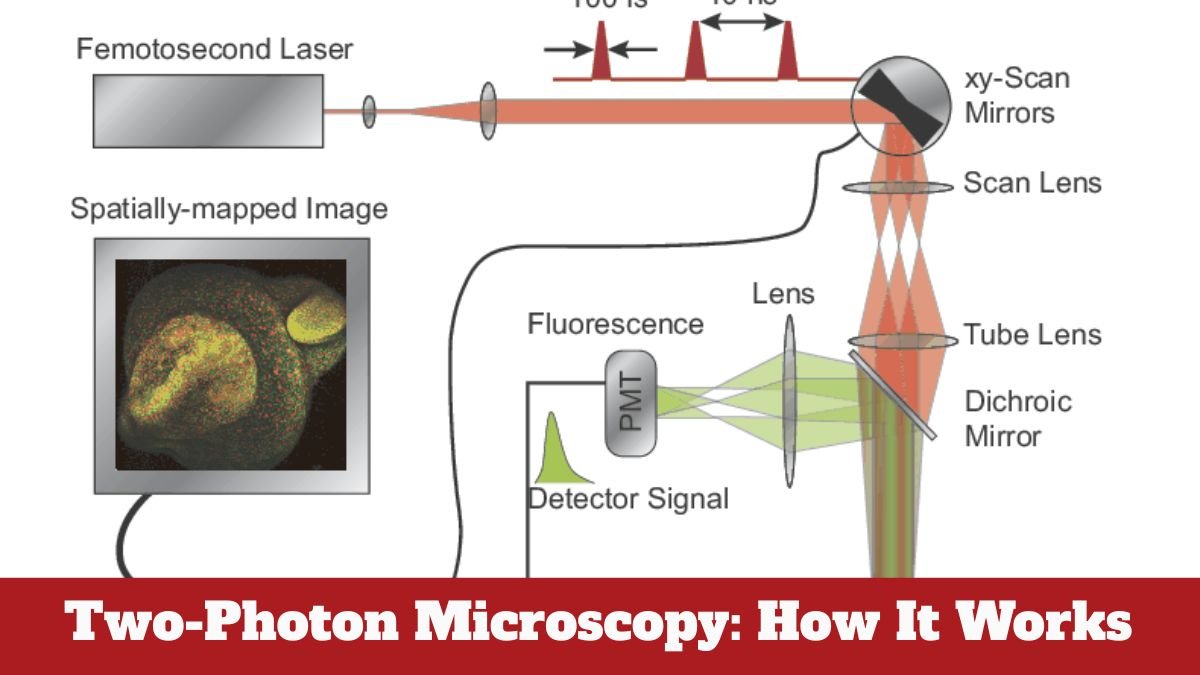
The Science of Two-Photon Excitation – How it Really Works
The fundamental principle of two-photon microscopy is two-photon excitation. In this process, two low-energy photons simultaneously excite a fluorophore. As the energy of both photons combines to form a higher energy, the fluorophore glows, creating an image. This process occurs only at the focal point, meaning the fluorescence is produced where the laser is focused. This reduces background noise and results in a clearer image.
How a Laser Creates Super-Detailed Images
Two-photon microscopy uses a special type of laser called a femto-second pulsed laser. This laser emits very short, yet intense pulses. These pulses contain enough photons to simultaneously excite the fluorophore. Since the excitation occurs only at the focal point, there is no damage to the surrounding area, and the image is produced with extremely high resolution.
Ability to See Deep in Tissue – Beyond Traditional Techniques
Traditional fluorescence microscopy can only see to a depth of 50–100 micrometers, but two-photon microscopy can easily image tissue 500–1000 micrometers (up to 1 mm). This makes it an excellent choice for brain imaging, tumor studies, and live animal imaging. Due to low scattering and reduced loss, this technique maintains clarity even at depth.
Why Tissue Damage Is Reduced – The Real Reason
The photons used in two-photon microscopy are lower in energy. This means the sample does not have to endure as much energy. Instead of harmful UV or high-energy light, near-infrared (NIR) light is used, which heats the sample less, causes less phototoxicity, and preserves the natural behavior of cells. This technique becomes a safer option for live cell imaging.
Live Cell Imaging – A Real Game Changer
The greatest advantage of two-photon microscopy is its ability to understand live cells in their natural environment. It allows real-time observation of cell-to-cell interactions, such as the activity of neurons in the brain, the movement of immune cells, or the proliferation of cancer cells. This was difficult with conventional microscopy because intense light would damage cells quickly. Two-photon technology has simplified this challenge.
How this technology is revolutionizing research – Uses and Importance
Two-photon microscopy is being used in many areas of modern science. In neuroscience, it helps understand brain circuits. In immunology, it records the activity of immune cells. In developmental biology, it helps observe the early stages of an embryo. This technology is providing researchers with more accurate, deep, and natural imaging than ever before.
Limitations and Challenges – Not Perfect, But Quite Powerful
Although two-photon microscopy is highly effective, it is not completely problem-free. It requires expensive lasers and high-quality optical systems. Its cost is quite high, making it difficult for small labs or institutions to afford. Furthermore, imaging speeds can sometimes be slow, especially when scanning large areas. However, its advantages far outweigh these challenges.
Two-Photon Microscopy in the Future – What Changes Ahead?
This technology is poised for further improvement in the future. With the advent of new lasers, better detectors, and faster scanning systems, this technology will become more rapid, affordable, and accessible. Multi-Photon Microscopy
Conclusion
Two-photon microscopy is a cutting-edge technology that has revolutionized the world of biological research. It offers remarkable breakthroughs in deep tissue visualization, live cell imaging with minimal damage, and the production of extremely clear images. Two-photon technology continues where traditional microscopy left off. This is why it is often called the future of scientific research.
